broncial tree drawing public domain
Gross Anatomy
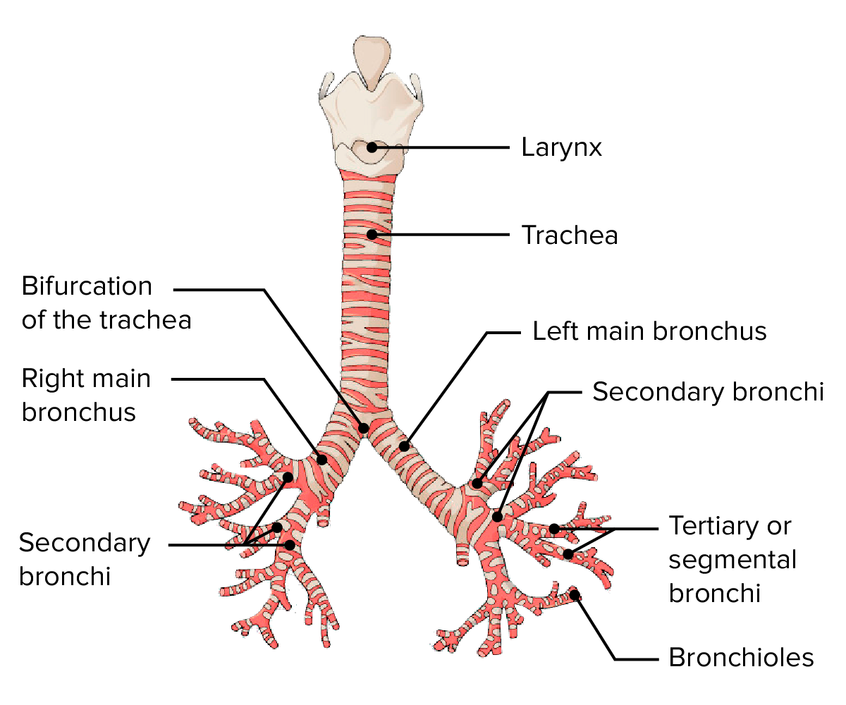
The bronchial tree begins at the bifurcation of the trachea Trachea The trachea is a tubular structure that forms part of the lower respiratory tract. The trachea is continuous superiorly with the larynx and inferiorly becomes the bronchial tree within the lungs. The trachea consists of a support frame of semicircular, or C-shaped, rings made out of hyaline cartilage and reinforced by collagenous connective tissue. Trachea: Anatomy at the carina, approximately at the level of T5. The trachea Trachea The trachea is a tubular structure that forms part of the lower respiratory tract. The trachea is continuous superiorly with the larynx and inferiorly becomes the bronchial tree within the lungs. The trachea consists of a support frame of semicircular, or C-shaped, rings made out of hyaline cartilage and reinforced by collagenous connective tissue. Trachea: Anatomy bifurcates into the main left and right bronchi, These bronchi continue to branch until they form alveoli Alveoli Small polyhedral outpouchings along the walls of the alveolar sacs, alveolar ducts and terminal bronchioles through the walls of which gas exchange between alveolar air and pulmonary capillary blood takes place. Acute Respiratory Distress Syndrome (ARDS) , the site of gas exchange Gas exchange Human cells are primarily reliant on aerobic metabolism. The respiratory system is involved in pulmonary ventilation and external respiration, while the circulatory system is responsible for transport and internal respiration. Pulmonary ventilation (breathing) represents movement of air into and out of the lungs. External respiration, or gas exchange, is represented by the O2 and CO2 exchange between the lungs and the blood. Gas Exchange . Each bronchial segment progressively becomes smaller in diameter and has a thinner wall.
Main branching structure and components
- Trachea Trachea The trachea is a tubular structure that forms part of the lower respiratory tract. The trachea is continuous superiorly with the larynx and inferiorly becomes the bronchial tree within the lungs. The trachea consists of a support frame of semicircular, or C-shaped, rings made out of hyaline cartilage and reinforced by collagenous connective tissue. Trachea: Anatomy → carina → main bronchi → lobar bronchi → segmental bronchi → terminal bronchioles → respiratory bronchioles → alveolar duct → alveolar sac → alveoli Alveoli Small polyhedral outpouchings along the walls of the alveolar sacs, alveolar ducts and terminal bronchioles through the walls of which gas exchange between alveolar air and pulmonary capillary blood takes place. Acute Respiratory Distress Syndrome (ARDS)
- Bronchi:
- Main bronchi:
- The left main bronchus is longer than the right and enters the left lung at the level of T6.
- The right main bronchus is wider, shorter, and more vertical than the left (most frequent pathway for aspirated foreign objects) and enters the right lung at the level of T5.
- Lobar or secondary bronchi:
- 2 left lobar bronchi
- 3 right lobar bronchi
- Segmental or tertiary bronchi:
- Each supplies a bronchopulmonary segment, which is the largest subdivision of a lobe.
- 10 segments in the right lung
- 8–10 segments in the left lung
- Main bronchi:
- Conducting bronchioles:
- 20–25 generations of branching
- End as terminal bronchioles
- Lack cartilage Cartilage Cartilage is a type of connective tissue derived from embryonic mesenchyme that is responsible for structural support, resilience, and the smoothness of physical actions. Perichondrium (connective tissue membrane surrounding cartilage) compensates for the absence of vasculature in cartilage by providing nutrition and support. Cartilage: Histology , glands, and alveoli Alveoli Small polyhedral outpouchings along the walls of the alveolar sacs, alveolar ducts and terminal bronchioles through the walls of which gas exchange between alveolar air and pulmonary capillary blood takes place. Acute Respiratory Distress Syndrome (ARDS) (distinguishing feature from bronchi)
- Terminal bronchioles give rise to several respiratory bronchioles.
- Respiratory bronchioles:
- Characterized by the outpocketing from their lumen: alveoli Alveoli Small polyhedral outpouchings along the walls of the alveolar sacs, alveolar ducts and terminal bronchioles through the walls of which gas exchange between alveolar air and pulmonary capillary blood takes place. Acute Respiratory Distress Syndrome (ARDS)
- Each respiratory bronchiole gives rise to 2–11 alveolar ducts.
- Each alveolar duct gives rise to 5–6 alveolar sacs, the site of gas exchange Gas exchange Human cells are primarily reliant on aerobic metabolism. The respiratory system is involved in pulmonary ventilation and external respiration, while the circulatory system is responsible for transport and internal respiration. Pulmonary ventilation (breathing) represents movement of air into and out of the lungs. External respiration, or gas exchange, is represented by the O2 and CO2 exchange between the lungs and the blood. Gas Exchange .

Anterior view of the larynx Larynx The larynx, also commonly called the voice box, is a cylindrical space located in the neck at the level of the C3-C6 vertebrae. The major structures forming the framework of the larynx are the thyroid cartilage, cricoid cartilage, and epiglottis. The larynx serves to produce sound (phonation), conducts air to the trachea, and prevents large molecules from reaching the lungs. Larynx: Anatomy , trachea Trachea The trachea is a tubular structure that forms part of the lower respiratory tract. The trachea is continuous superiorly with the larynx and inferiorly becomes the bronchial tree within the lungs. The trachea consists of a support frame of semicircular, or C-shaped, rings made out of hyaline cartilage and reinforced by collagenous connective tissue. Trachea: Anatomy , and bronchial tree:
Note the main divisions of the bronchi.
Neurovasculature
Blood supply:
The bronchial tree is supplied by branches of the left and right bronchial arteries Arteries Arteries are tubular collections of cells that transport oxygenated blood and nutrients from the heart to the tissues of the body. The blood passes through the arteries in order of decreasing luminal diameter, starting in the largest artery (the aorta) and ending in the small arterioles. Arteries are classified into 3 types: large elastic arteries, medium muscular arteries, and small arteries and arterioles. Arteries: Histology .
- Left bronchial artery: a direct branch of the thoracic aorta Aorta The main trunk of the systemic arteries. Mediastinum and Great Vessels: Anatomy
- Right bronchial artery: varied origin: branch of the thoracic aorta Aorta The main trunk of the systemic arteries. Mediastinum and Great Vessels: Anatomy , an intercostal artery on the right side, or one of the superior branches of the left bronchial artery
Venous drainage:
- The bronchial veins Veins Veins are tubular collections of cells, which transport deoxygenated blood and waste from the capillary beds back to the heart. Veins are classified into 3 types: small veins/venules, medium veins, and large veins. Each type contains 3 primary layers: tunica intima, tunica media, and tunica adventitia. Veins: Histology are analogous to the bronchial arteries Arteries Arteries are tubular collections of cells that transport oxygenated blood and nutrients from the heart to the tissues of the body. The blood passes through the arteries in order of decreasing luminal diameter, starting in the largest artery (the aorta) and ending in the small arterioles. Arteries are classified into 3 types: large elastic arteries, medium muscular arteries, and small arteries and arterioles. Arteries: Histology .
- The bronchial tree is drained by tributaries of the main left and right bronchial veins Veins Veins are tubular collections of cells, which transport deoxygenated blood and waste from the capillary beds back to the heart. Veins are classified into 3 types: small veins/venules, medium veins, and large veins. Each type contains 3 primary layers: tunica intima, tunica media, and tunica adventitia. Veins: Histology :
- Left bronchial vein drains into the left superior intercostal vein or the accessory hemiazygos vein.
- Right bronchial vein drains into the azygos vein.
Innervation:
Innervation is supplied by the pulmonary plexus of the vagus nerve Vagus nerve The 10th cranial nerve. The vagus is a mixed nerve which contains somatic afferents (from skin in back of the ear and the external auditory meatus), visceral afferents (from the pharynx, larynx, thorax, and abdomen), parasympathetic efferents (to the thorax and abdomen), and efferents to striated muscle (of the larynx and pharynx). Pharynx: Anatomy .
Source: https://www.lecturio.com/concepts/bronchial-tree/
0 Response to "broncial tree drawing public domain"
Post a Comment